
Recently, a research team led by Prof. WANG Junfeng at the High Magnetic Field Laboratory, the Hefei Institutes of Physical Science of the Chinese Academy of Sciences, has developed an innovative biomimetic dual-mode magnetic resonance imaging (MRI) nanoprobe for detecting early-stage liver fibrosis in non-alcoholic fatty liver disease (NAFLD).
The work, using the Steady High Magnetic Field Facility (SHMFF), was recently published in Advanced Science.
NAFLD is a growing global health concern with even higher rates among individuals with obesity or type 2 diabetes. Detecting liver fibrosis early, before it becomes irreversible, is crucial for timely intervention and treatment. While MRI is a promising non-invasive tool for identifying liver fibrosis, traditional imaging techniques often lack the sensitivity to catch early-stage changes. Conventional contrast agents either face safety concerns or fail to target fibrotic tissues specifically.
In this study, the researchers developed a new type of nanoprobe that mimics natural biological processes—specifically, protein biomineralization—and targets key biomarkers of early fibrosis, such as PDGFRβ, a receptor overexpressed by activated liver cells involved in fibrosis development.
Using biomineralized bovine serum albumin (BSA) as a framework, the team created a dual-mode contrast agent capable of enhancing both T1 and T2 MRI signals. This approach combines the strengths of two imaging modes: T1-weighted images highlight fibrotic lesions, while T2-weighted images suppress background noise, providing clearer, more accurate results.
In laboratory and cellular tests, the nanoprobe demonstrated high imaging sensitivity, specific targeting of fibrotic cells, and excellent biocompatibility. When tested using a 7 Tesla MRI system, the nanoprobe enabled precise visualization of early-stage fibrosis within just one hour, significantly improving diagnostic speed and accuracy.
This work provides a precise diagnostic tool for early liver fibrosis and holds significant clinical potential for disease prognosis assessment and recurrence monitoring.
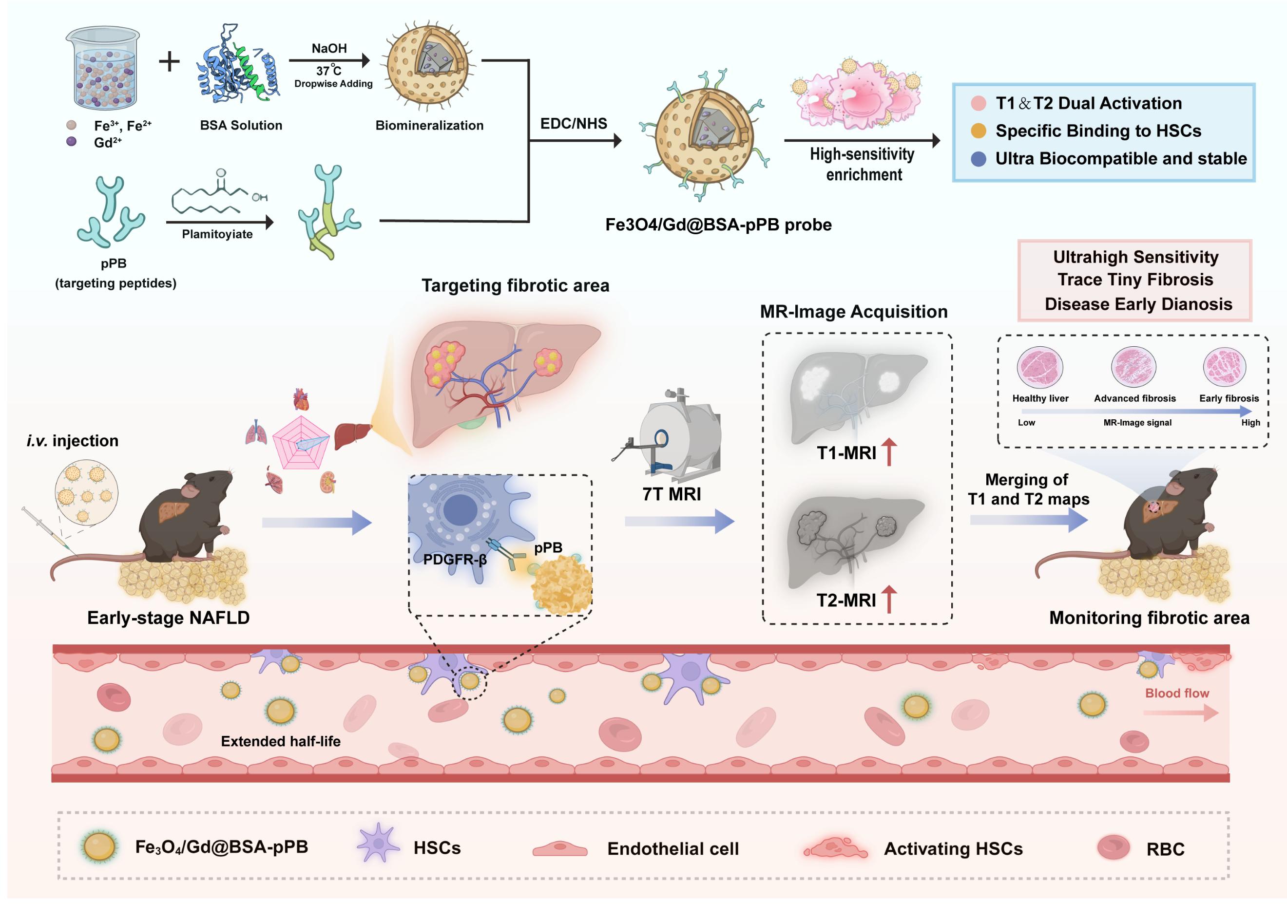
Early Diagnosis of Liver Fibrosis Using Dual-Mode T1 and T2 Imaging with the Fe₃O₄/Gd@BSA-pPB Nanoprobe (Image by MA Kun)
![Schematic illustration of (a) the preparation of Mn3[Co(CN)6]2@SiO2@Ag and (b) the delivery process of these Dox-loaded Mn3[Co(CN)6]2@SiO2@Ag to A549 cells for enhanced fluorescence imaging](../../../news/Events/201601/W020160122330206981460.png)