
Recently, a team led by Prof. HUANG Qing at the Institute of Intelligent Machines, Hefei Institutes of Physical Science (HFIPS) constructed a novel biosensor based on surface-enhanced Raman spectroscopy (SERS) to detect inflammatory Interleukin-6 (IL-6) protein in serum samples.
In addition, the aptamer-SERS detection protocols they developed can detect environmentally hazardous substances such as coralyne, PCB-77 and Hg2+.
In this research, scientists fabricated highly sensitive and specific aptamer-functionalized Au nanoparticles array, and quantitatively measured Raman signals from IL-6 protein in mice serum as low as 0.8 pM in 10-12-10-7 M concentration range. Besides, the researchers demonstrated the applicability of SERS biosensors by detecting the IL-6 in radiation injury, drug and bacterial infection cases by analyzing the serum of infected mice.
Researchers also categorized the aptamer-SERS biosensors into two categories in a review article for better understanding, in which label-free biosensors based on intrinsic conformation change of aptamer, and Raman reporter labeled aptamer-SERS biosensors were listed. The latter were further divided into three sub-types basing on “on/off approach”, “sandwich” approach, and catalytic approach. It showed the schematic diagrams with extensive examples to elaborate the working strategies and analytical applications.
The review as a whole highlighted the respective advantages, shortcomings, and available opportunities for the aptamer-SERS technique, which may help researchers to further develop the aptamer-based biosensors and make them useful in practical applications such as detecting disease specific and inflammatory biomarkers.
This research was supported by the National Natural Science Foundation of China and China Scholarship Council.
Link to the paper: Aptamer-functionalized Au nanoparticles array as the effective SERS biosensor for label-free detection of interleukin-6 in serum
A review of aptamer-based SERS biosensors: Design strategies and applications
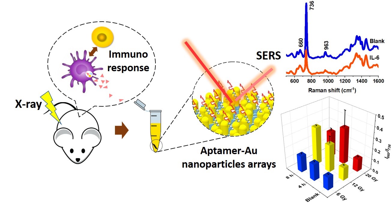
Fig.1. Schematic plot for SERS detection of IL-6 in serum of mice exposed to X-ray irradiation. (Image by Muhammad)
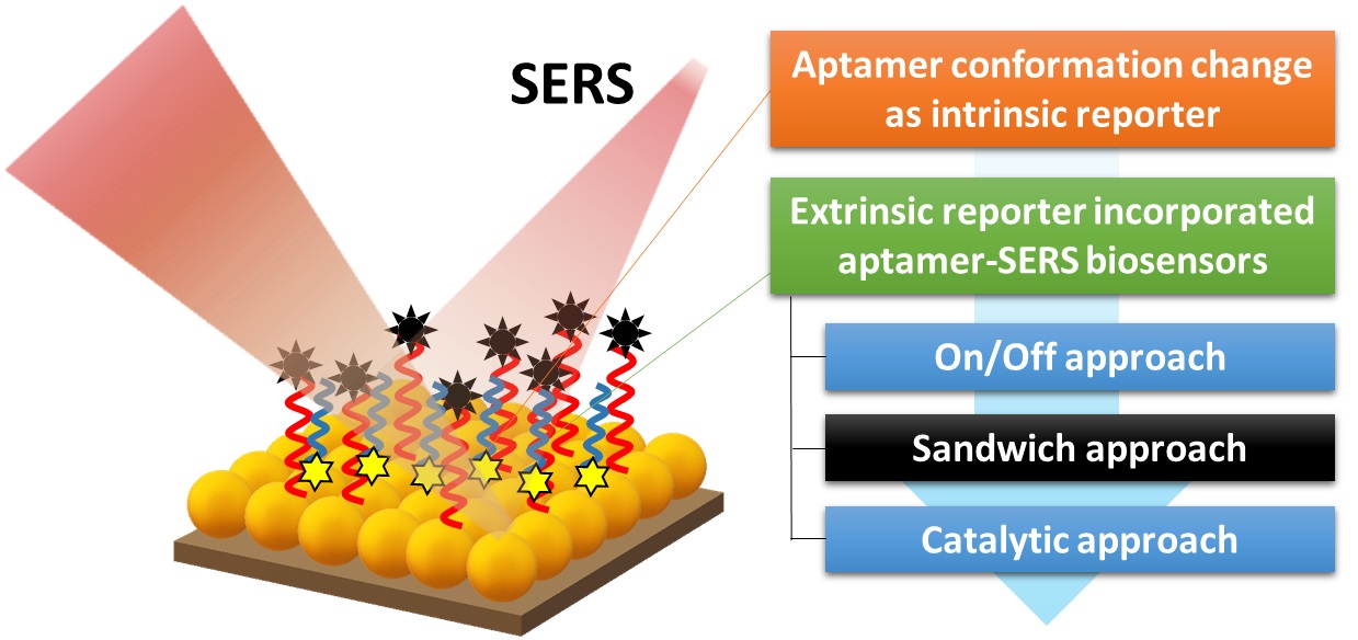
Fig.2. Review of major aptamer-SERS protocols. (Image by Muhammad)

Fig. 3. Schematic illustration for the detection of (A) Hg2+, (B) PCB-77, (C) Coralyne, using aptamer-SERS biosensors. (Image by Muhammad)
Contact:
ZHAO Weiwei
Hefei Institutes of Physical Science (http://english.hf.cas.cn/)
Email: annyzhao@ipp.ac.cn