
Using the shrink-assembled liquid 3D hot spot matrix as a microreactor, a new method for quantitative detection of blood drug concentration by surface-enhanced Raman spectroscopy (SERS) was established by a collaborated research team led by Prof. YANG Liangbao and Prof. WANG Hongzhi from Hefei Institutes of Physical Science (HFIPS), Chinese Academy of Sciences (CAS), with advantages of high stability and high sensitivity.
The results were published in Analytical Chemistry recently.
Quantitative detection is one of the ultimate goals of SERS methods, but there have been challenges in controlling the homogeneity of the hot spot and getting target molecules into the hot spot region.
Profesor YANG Liangbao's team has been engaged in the research work on the principle and detection application of SERS method for years, which is a type of fast, highly sensitive, and fingerprinted molecular spectroscopy.
A series of achievements have been made with SERS in their previous work, including detection of poisons in oily matrix, and actively capturing target molecules in small gaps based on nano-capillary pumping model.
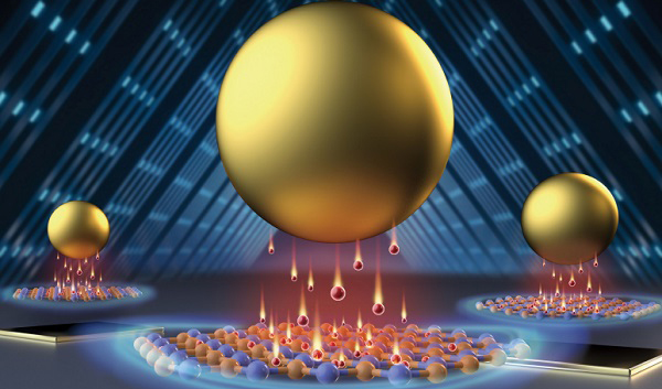
In this work, scientists used the liquid three-phase equilibrium principle to control the shrinkage of droplets, which not only forms a high-density and highly stable liquid 3D hotspot matrix, but also enables antitumor drugs to enter the hotspot region autonomously. Then with a self-made effectively hand-held Raman spectrometer, they realized, for the first time, the online quantitative detection of anticancer drugs in the serum of tumor patients.
The method exhibited 50ppb sensitivity and a quantitative detection range of 50-1000ppb for the anticancer drug 5-fluorouracil.
Compared with traditional solid nanoarrays and colloidal aggregation SERS methods, the shrink-assembled liquid 3D hotspot matrix can enhance the enrichment ability of analytes in the plasmonic hotspot space, enabling highly sensitive and highly stable SERS quantitative detection.
"It has great potential for quantitative detection of analytes in complex samples like serum, biological fluids, dynamic monitoring of anticancer drug metabolism processes and biochemical reaction kinetics," said Prof. YANG.
This research was funded by the Scientific Research Instrument and Equipment Development Project of the Chinese Academy of Sciences (YJKYYQ20200022), the National Natural Science Foundation of China (No. 21974142), and the Key R&D Program of Anhui Province (No. 202104d07020002).
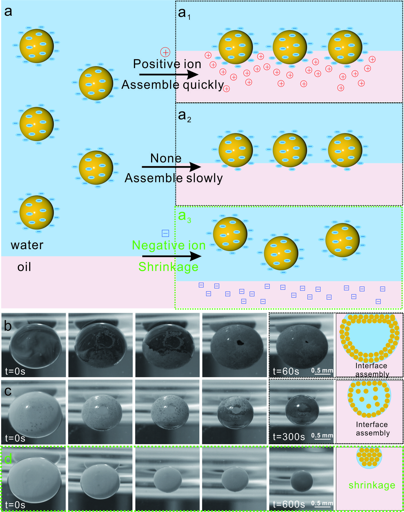
Figure 1. Schematic diagram of 3D hotspot matrix formation. (Image by ZHOU Guoliang)

Figure 2. Detection of 5-fluorouracil in serum by hand-held Raman spectrometer. (Image by ZHOU Guoliang)