Recently, a joint team from Hefei Institutes of Physical Science (HFIPS) of Chinese Academy of Sciences (CAS) has made new progress in the research of "radiotherapy brain".
The related results were published in the Journal of Magnetic Resonance Imaging.
Professor Lauren J. O'Donnell, a neuroimaging specialist at Harvard Medical School, wrote an editorial commentary on behalf of the journal's editorial board, describing the study as an innovative approach to quantitative tract-based analysis that offers a promising idea for future "radiation therapy brain" research.
Radiotherapy is an essential therapy for brain tumors, and two critical factors should be considered in radiotherapy planning: radiotherapy effect and brain function protection.
To achieve the optimal quality of radiotherapy, Hefei Cancer Hospital of the Chinese Academy of Sciences has formed a joint research team of radiotherapy, imaging, cognitive and clinical experts since 2017, which is dedicated to the early assessment, intervention of precise radiotherapy and the risk of late radiation brain injury.
Since the start of the project, the joint team has taken patients with nasopharyngeal carcinoma as the primary research target and adequately protected the hippocampus. And the MRI neuroimaging data and neurocognitive data of patients were also collected before, during, and after radiotherapy to track the effect of radiotherapy in real-time and assess the quality of radiotherapy, to establish an early prediction model for the risk of late-onset radiation brain injury and providing a technical means to maximize the quality of patient treatment.
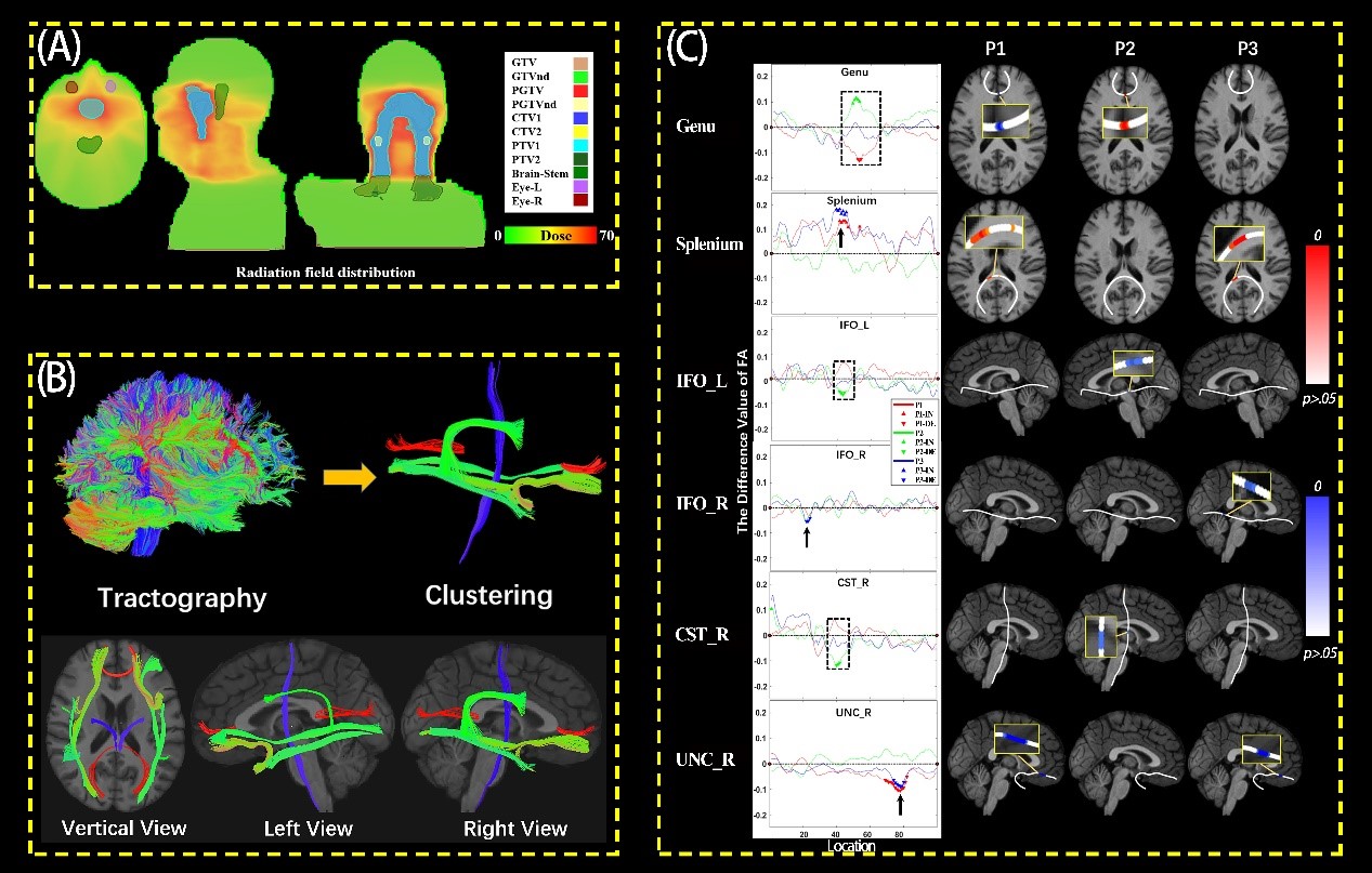
In 2021, based on some preliminary data, the team used a tract-based analysis to explore the sensitivity of radiotherapy to the significant nerve fiber bundles in the brain of patients with nasopharyngeal carcinoma, laying the foundation for future personalized radiotherapy.

Microscopic morphological changes of white matter fiber bundles during radiotherapy. (Image by YANG Lizhuang)