
Recently, researchers from the Hefei Institutes of Physical Science of the Chinese Academy of Sciences, successfully constructed a three-dimensional(3D) lung cell model. They used solid phase microextraction-gas chromatography-mass spectrometry (SPME-GC-MS) non-targeted assay to obtained differential volatile organic compounds (VOCs) released by lung cancer and lung epithelial cells in both two-dimensional(2D) and (three-dimensional) 3D states.
"Understanding how cells behave in different environments could help us learn more about diseases like cancer and how to treat them," said HUANG Ying, memeber of the team.
The findings were published as the cover article in the Journal of Chinese Mass Spectrometry Society.
VOCs released by cancer cell metabolism have been extensively researched as a potential tumor marker. However, the majority of in vitro cell culture utilizes adherent 2D growth mode, which differs from the 3D structure of tumors in the human body.
In this study, researchers examined how lung cancer cells (A549) and normal lung epithelial cells (BEAS-2B) behave in different environments. They grew these cells in two ways: the usual flat, 2D way and a newer, more realistic 3D method called the hanging drop technique. With SPME-GC-MS technique, they found that the release of hydrocarbons (alcohols, acids, alkanes) and proline metabolites (pyrroline) in 3D cell spheres increased by 2-13 times compared to 2D cells. This indicates that oxidative stress response and proline metabolism were more active in 3D cell spheres.
The in vitro cell 3D culture method established in this study will provide a new technical approach for studying cancer VOCs markers and their formation mechanism.
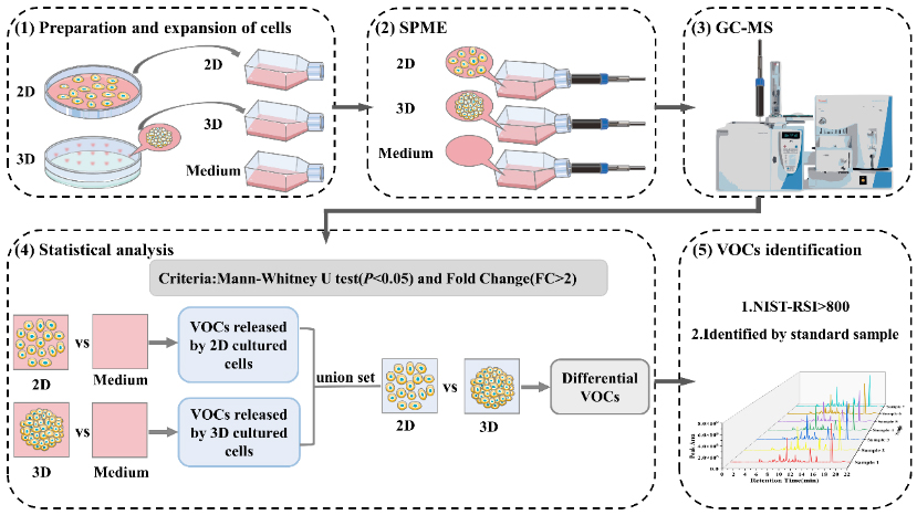
Cell Experiment and Assay Analysis (Image by HUANG Ying)