A research team led by Prof. CHU Yannan from the Hefei Institutes of Physical Science of the Chinese Academy of Sciences, together with collaborators in China, the United States and Singapore, has developed an artificial intelligence model to improve the classification of pulmonary nodules on chest CT images.
The study was published in Information Fusion.
Distinguishing benign and malignant pulmonary nodules is important for early lung cancer detection. In clinical practice, radiologists usually review CT images from an overall view before focusing on the detailed features of individual nodules. This process requires careful analysis and can be time-consuming.
In this study, the researchers developed M3Net, a three-dimensional AI model that analyzes CT images at different scales.
Inspired by radiologists' reading process, the model examines information from the overall lung structure, the area surrounding nodules, and details within the nodules. By combining information from the nodule itself and the surrounding lung tissue, M³Net can identify features associated with malignancy.
The team evaluated M3Net using the public LIDC-IDRI dataset and a clinical dataset from the University of Science and Technology of China. Compared with 12 existing AI methods, M³Net achieved classification accuracies of 86.96% and 84.24% on the two datasets, respectively, showing higher accuracy than the best-performing comparison method.
The researchers also tested the model on challenging cases, including nodules with unclear boundaries or unusual appearances. Visualization analysis showed that M³Net focused on regions related to diagnosis, helping researchers better understand the basis of its predictions.
By incorporating a radiologist-like hierarchical approach into AI-based image analysis, M³Net may provide support for pulmonary nodule assessment and computer-aided diagnosis.
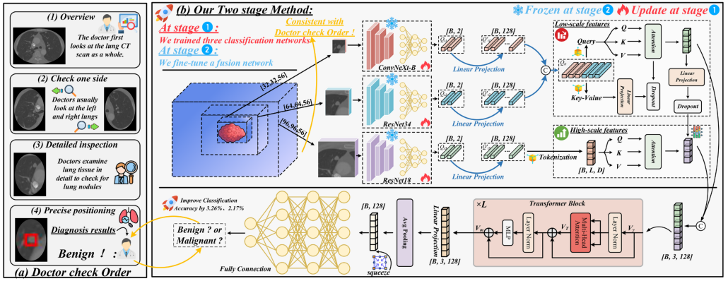
Workflow of the clinically inspired M3Net model for pulmonary nodule classification (Image by GE Dianlong)
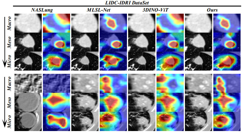
Grad-CAM comparison of different models using multi-scale pulmonary nodule CT images (Image by GE Dianlong)