
A research team led by Professor LI Hai from the Hefei Institutes of Physical Science of the Chinese Academy of Sciences (CAS), has for the first time demonstrated that combining surface-based morphometry (SBM) with cortical diffusion tensor imaging (DTI) can sensitively detect early structural changes in the temporal lobe of nasopharyngeal carcinoma (NPC) patients during radiotherapy.
Their findings were recently published in the Journal of Magnetic Resonance Imaging.
Radiotherapy is the primary treatment for NPC, but because the temporal lobe lies close to the nasopharynx, it is particularly vulnerable to radiation-induced damage. Detecting subtle structural changes at an early stage is therefore critical for both treatment precision and the protection of brain function.
In this study, 40 NPC patients underwent Magnetic Resonance Imaging (MRI) scans at three points—before, during, and after conventional radiotherapy. By combining SBM and cortical DTI analyses, the researchers evaluated changes in two imaging indicators: cortical thickness and cortical mean diffusivity. This dual-perspective approach, integrating both macroscopic morphology and microscopic diffusional changes, was shown to improve sensitivity in detecting temporal lobe alterations. Importantly, the results also highlighted how these indicators complement each other, helping to reveal both radiation sensitivity and the relationship between dose and brain response.
The research is part of the long-term efforts of the "radiotherapy brain" joint team, established in 2017 by Hefei Cancer Hospital, CAS. Bringing together specialists in radiotherapy, medical imaging, and cognitive science, the team has been dedicated to developing strategies for precision radiotherapy and for assessing and preventing delayed radiation-induced brain injury. In 2022, they applied fiber bundle-based analysis to study radiation sensitivity in major white matter tracts.
This new study has enriched the understanding of radiation effects on the brain, providing new insights that may support personalized precision radiotherapy for NPC patients, according to the team.
The research was supported by grants from the National Natural Science Foundation of China, the Anhui Provincial Natural Science Foundation, and key R&D projects in Anhui Province.
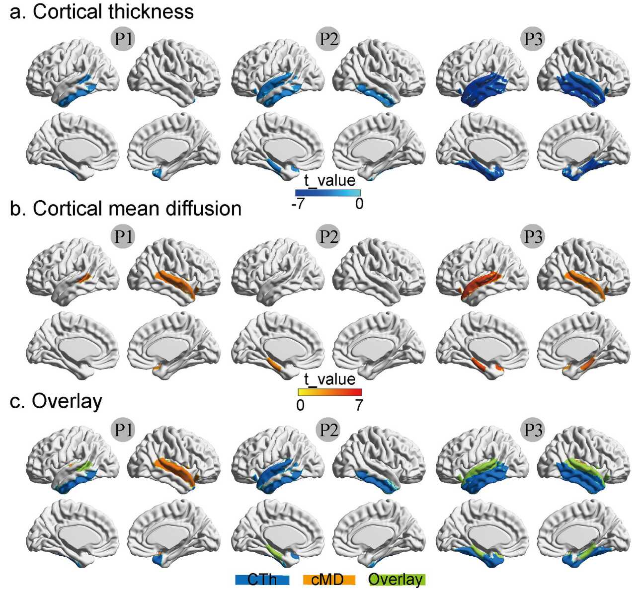
Early Onset of Microstructural Changes During Radiation Therapy Sessions. (Image by YANG Lizhuang)